Home » Without Label » Diagram Of Backbone : lumbar-spine-diagram-labeled-human-backbone-keywords-a ... / It is made up of 33 bones, known as vertebra (plural ~ vertebrae), which may be fused at certain points, like in.
Diagram Of Backbone : lumbar-spine-diagram-labeled-human-backbone-keywords-a ... / It is made up of 33 bones, known as vertebra (plural ~ vertebrae), which may be fused at certain points, like in.
Diagram Of Backbone : lumbar-spine-diagram-labeled-human-backbone-keywords-a ... / It is made up of 33 bones, known as vertebra (plural ~ vertebrae), which may be fused at certain points, like in.. The fifth lumbar spine vertebrae (l5) is part of the greater lumbar region. The intervertebral foramen (neural passageways. The thoracic spine helps keep the body upright and stable. Certain back muscles extend to other areas, like the shoulders, upper arms, and thighs. On the chart below you will see 4 columns (vertebral level, nerve root, innervation, and possible symptoms).
We hope this picture anatomy of back muscles diagram can help you study and research. Human anatomy · july 23, 2016. The atlas is the topmost vertebra, and along with c2, forms the joint connecting the skull and spine. The spine anatomy is a complex structure. We think this is the most useful anatomy picture that you need.
Vertebral Column Anatomy Poster | Spine Anatomical Chart ... from www.anatomystuff.co.uk The thoracic spine helps keep the body upright and stable. See lumbar spine anatomy diagram stock video clips. The vertebral column is the most important collection of bones to maintain stability of the skeletal structure and support of the entire body, especially when upright. The vertebrae of the spine align so that their vertebral canals form a hollow, bony tube to protect the spinal cord from external damage and infection. The intervertebral foramen (neural passageways. Anatomynote.com found anatomy of back muscles diagram from plenty of anatomical pictures on the internet. The fifth lumbar spine vertebrae (l5) is part of the greater lumbar region. As you can see in the vertebrae diagram above, the human spine consists of 33 vertebrae in total;
Human backbone diagram, bone, human backbone diagram.
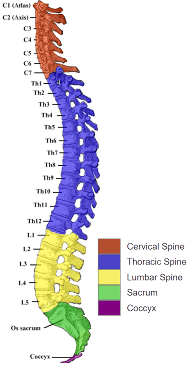
Studying a spine diagram is one way to better understand many of the individual components of the back bone and how they might relate to a symptomatic back, neck or sciatica pain condition. Anatomynote.com found anatomy of back muscles diagram from plenty of anatomical pictures on the internet. Spine diagrams the human spine consists of 33 vertebrae: The thoracic spine is the middle part of the spine, connecting the cervical and lumbar spine. Simply line up the vertebral level with the possible symptoms and you will see some surprising connections of symptoms that relate to your spine. The trapezius or trapezoid muscles are two paired muscles that extend from the base of the thoracic vertebrae in the spine to the occipital bone and run out to the spine of the scapula. On the chart below you will see 4 columns (vertebral level, nerve root, innervation, and possible symptoms). Spinal anatomy is a remarkable combination of strong bones, flexible ligaments and tendons, large muscles and highly sensitive nerves. Between the vertebrae are small spaces known as intervertebral canals that allow spinal nerves to exit the spinal cord and connect to the various regions of the body. Your spine, or backbone, is your body's central support structure. It is made up of 33 bones, known as vertebra (plural ~ vertebrae), which may be fused at certain points, like in. The intervertebral foramen (neural passageways. 24 are considered to be part of the upper spine, whilst the other 11 are found in the sacrum & coccyx.
We hope this picture anatomy of back muscles diagram can help you study and research. Backbone is jquery's best friend so to speak so you 'organize' your code and use jquery to query the dom. 12 photos of the human back bone chart. Your spine, or backbone, is your body's central support structure. The intervertebral foramen (neural passageways.
Human Spine and Spinal Cord Picture C1 - S5 Vertebra ... from www.disabled-world.com The vertebral column of the lower back includes the five lumbar vertebrae, the sacrum, and the coccyx. Anatomy of the back organs. The thoracic spine helps keep the body upright and stable. Made up of 34 bones, the spinal column holds the body upright, allows it to bend and twist with ease and provides a conduit for major nerves running from the brain to the tips of the toes—and everywhere in between. Your spine, or backbone, is your body's central support structure. The back functions are many, such as to house and protect the spinal cord, hold the body and head upright, and adjust the movements of the upper and lower limbs. The thoracic spine is the middle part of the spine, connecting the cervical and lumbar spine. For more anatomy content please follow us and visit our website:
The vertebral column of the lower back includes the five lumbar vertebrae, the sacrum, and the coccyx.
Made up of 34 bones, the spinal column holds the body upright, allows it to bend and twist with ease and provides a conduit for major nerves running from the brain to the tips of the toes—and everywhere in between. It happens as your body ages, but it also can. It is designed to be incredibly strong, protecting the highly sensitive nerve roots, yet highly flexible, providing for mobility on many different planes. The vertebrae of the spine align so that their vertebral canals form a hollow, bony tube to protect the spinal cord from external damage and infection. Spinal anatomy is a remarkable combination of strong bones, flexible ligaments and tendons, large muscles and highly sensitive nerves. Your spine, or backbone, is your body's central support structure. The spinous processes are horizontal and more squared in shape. Certain back muscles extend to other areas, like the shoulders, upper arms, and thighs. These vertebrae bear much of the body's weight and related biomechanical stress. Spondylolisthesis is a main cause of lower back pain. Human anatomy · july 23, 2016. Simply line up the vertebral level with the possible symptoms and you will see some surprising connections of symptoms that relate to your spine. It comprises the vertebral column (spine) and two compartments of back muscles;
Bones of the pelvis and lower back. Anatomynote.com found anatomy of back muscles diagram from plenty of anatomical pictures on the internet. For more anatomy content please follow us and visit our website: We hope this picture anatomy of back muscles diagram can help you study and research. See lumbar spine anatomy diagram stock video clips.
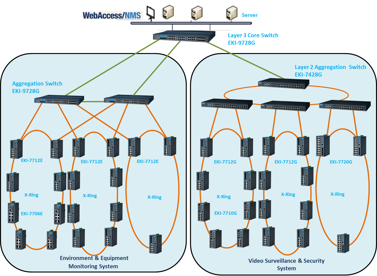
Building an Effective Ethernet Backbone Network for China ... from advcloudfiles.advantech.com The spine diagram shown below, consists of many bones or vertebrae,soft discs,the spinal cord, and spinal nerves. Backbone is jquery's best friend so to speak so you 'organize' your code and use jquery to query the dom. The atlas is the topmost vertebra, and along with c2, forms the joint connecting the skull and spine. The lower part of the trapezius ascends and depresses the scapula, while the transverse or middle region of the trapezius is what retracts the. The trapezius or trapezoid muscles are two paired muscles that extend from the base of the thoracic vertebrae in the spine to the occipital bone and run out to the spine of the scapula. Studying a spine diagram is one way to better understand many of the individual components of the back bone and how they might relate to a symptomatic back, neck or sciatica pain condition. Anatomical diagrams of the spine and back this human anatomy module is composed of diagrams, illustrations and 3d views of the back, cervical, thoracic and lumbar spinal areas as well as the various vertebrae. The intervertebral foramen (neural passageways.
For more anatomy content please follow us and visit our website:
It is made up of 33 bones, known as vertebra (plural ~ vertebrae), which may be fused at certain points, like in. The thoracic spine is the middle part of the spine, connecting the cervical and lumbar spine. The spine diagram shown below, consists of many bones or vertebrae,soft discs,the spinal cord, and spinal nerves. The back is the body region between the neck and the gluteal regions. See lumbar spine anatomy diagram stock video clips. The bones of the pelvis and lower back work together to support the body's weight, anchor the abdominal and hip muscles, and protect the delicate vital organs of the vertebral and abdominopelvic cavities. These vertebrae bear much of the body's weight and related biomechanical stress. The thoracic spine helps keep the body upright and stable. Anatomical diagrams of the spine and back this human anatomy module is composed of diagrams, illustrations and 3d views of the back, cervical, thoracic and lumbar spinal areas as well as the various vertebrae. Backbone is jquery's best friend so to speak so you 'organize' your code and use jquery to query the dom. Human backbone diagram, bone, human backbone diagram. Spinal vertebrae bone spine vertebra toracica spinal cord spine structure back diagram spine sections spinal cord vertebrae spinal structure health diagram. Between the vertebrae are small spaces known as intervertebral canals that allow spinal nerves to exit the spinal cord and connect to the various regions of the body.